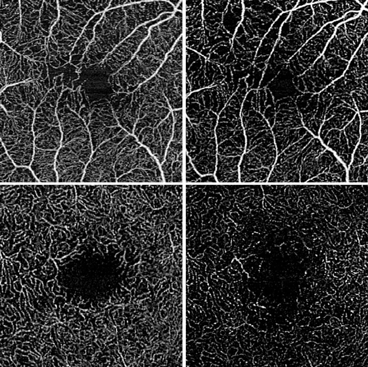
Optical coherence tomography angiography showing ocular vascular perfusion in intermediate uveitis:
The upper images show the superficial and the lower images the deep retinal blood flow. The initial state is shown on the left and the state after disease worsening is shown on the right. The blood flow density has decreased in both the superficial and deep retinal layers.
